Abstract
Prune Belly Syndrome (PBS) is a rare congenital disorder characterized by the classical triad of deficient abdominal wall musculature, urinary tract anomalies, and bilateral cryptorchidism in males. The condition is associated with significant morbidity due to urinary tract dysfunction, recurrent infections, and potential renal impairment. We report two siblings with PBS or a pseudo-PBS phenotype, highlighting severe urinary tract involvement and a possible familial predisposition. The first case was a preterm male neonate delivered at 34 weeks of gestation by cesarean section after antenatal ultrasonography demonstrated bilateral hydronephrosis. He subsequently developed recurrent vomiting, progressive abdominal distension, urinary retention, high-grade bilateral vesicoureteral reflux, and a neurogenic bladder. His clinical course was complicated by Burkholderia cepacia sepsis, thrombocytopenia, electrolyte disturbances, hyperammonemia, and respiratory distress requiring intensive care. Pediatric urology recommended vesicostomy and genetic evaluation. The second case was a female sibling with a prenatal diagnosis of a distended bladder and bilateral hydronephroureter who had a pseudo-PBS phenotype and developed severe urinary tract disease, recurrent infections, feeding intolerance, meningitis, and progressive renal failure, ultimately resulting in death at 3.9 years of age. These cases emphasize the importance of early recognition of PBS, particularly when antenatal urinary tract abnormalities and a positive family history are present, and highlight the need for multidisciplinary management, genetic counseling, and long-term renal and urological follow-up. A preterm male neonate was delivered at 34 weeks of gestation via cesarean section. Antenatal ultrasonography revealed bilateral hydronephrosis. The patient had a significant family history of a sibling who had died from pseudo-prune belly syndrome. Shortly after birth, he developed recurrent vomiting, progressive abdominal distension, and urinary retention. Radiological investigations demonstrated severe bilateral vesicoureteral reflux and a neurogenic bladder, confirming the diagnosis of PBS. During hospitalization, the infant developed sepsis caused by Burkholderia cepacia, accompanied by electrolyte disturbances and hyperammonemia, necessitating intensive care management. Treatment included broad-spectrum antibiotics, blood transfusions, ventilatory support, and close monitoring. Pediatric urology consultation recommended vesicostomy to relieve urinary obstruction and genetic testing to evaluate the possible familial basis of the disorder. This case highlights the importance of early recognition of PBS, particularly in neonates with antenatal urinary tract abnormalities and a positive family history. The occurrence of severe complications, including sepsis and metabolic derangements, underscores the need for prompt multidisciplinary intervention. Genetic counseling and evaluation should be considered in families with recurrent or related congenital anomalies to facilitate early diagnosis, guide management, and improve long-term outcomes.
|
Published in
|
Science Discovery Pediatrics (Volume 1, Issue 1)
|
|
DOI
|
10.11648/j.sdpediatr.20260101.12
|
|
Page(s)
|
13-18 |
|
Creative Commons
|

This is an Open Access article, distributed under the terms of the Creative Commons Attribution 4.0 International License (http://creativecommons.org/licenses/by/4.0/), which permits unrestricted use, distribution and reproduction in any medium or format, provided the original work is properly cited.
|
|
Copyright
|
Copyright © The Author(s), 2026. Published by Science Publishing Group
|
Keywords
Prune Belly Syndrome, Eagle–Barrett syndrome, Vesicoureteral Reflux, Hydronephrosis, Neurogenic Bladder
1. Introduction
Prune belly syndrome is a rare condition present at birth that involves problems that range from mild to severe. Prune belly syndrome is most often found in utero or at birth.
.
The condition has also been called the triad syndrome because of the three major features these include: weak or missing abdominal muscles, one or both undescended testicles, and an abnormally large bladder with kidney problems.
| [1] | Giorgi, A. (2024, April 22). Prune belly syndrome: What it is and what to expect. Verywell Health.
https://www.verywellhealth.com/prune-belly-syndrome-8622644 |
| [2] | Alkhamis, W. H., Abdulghani, S. H. & Altaki, A. Challenging diagnosis of prune belly syndrome antenatally: a case report. J Med Case Reports 13, 198 (2019).
https://doi.org/10.1186/s13256-019-2120-x |
| [13] | Snider Novitski K, Radulescu A, Jelin EB, Ladd MR, Meier K, Novotny NM. Prune Belly Syndrome. In: Hirschl RR, Powell DD, Waldhausen JJ, eds. Pediatric Surgery NaT. American Pediatric Surgical Association; 2023.
https://www.pedsurglibrary.com/apsa/view/Pediatric-Surgery-NaT/829694/all/Prune_Belly_Syndrome Accessed August 25, 2025 |
[1, 2, 13]
.
The exact aetiology of PBS is unknown, although several embryologic theories attempt to explain the anomal. But mostly, It is caused by urethral obstruction early in development resulting in massive bladder distension and urinary ascites, leading to degeneration of the abdominal wall musculature and failure of testicular descent. The impaired elimination of urine from the bladder leads to oligohydramnios, pulmonary hypoplasia, and Potter's facies.
| [6] | Metwalley, K. A., Farghalley, H. S. & Abd-Elsayed, A. A. Prune belly syndrome in an Egyptian infant with Down syndrome: A case report. J Med Case Reports 2, 322 (2008).
https://doi.org/10.1186/1752-1947-2-322 |
[6]
.
Symptoms vary since prune belly syndrome affects each person uniquely. It is common to have frequent UTIs, which can lead to kidney damage. Other anatomical anomalies can be associated with anomalies of the cardiorespiratory, skeletal, gastrointestinal, and central nervous system in 75% of the cases.
.
Diagnosis of PBS is clinical and radiological investigations show only the extent of renal dysplasia and/or renal disfunction.
| [10] | Kheir, A. E. M., Ali, E. M. A., Medani, S. A., & Maaty, H. S. (2017). Prune belly syndrome: A report of 15 cases from Sudan. Sudanese journal of paediatrics, 17(1), 42–48. |
[10]
.
Early diagnosis can help babies get the right treatment as soon as possible for the best results. The effects of prune belly syndrome are severe or deadly for some babies. However, many babies have minor defects and remain in good physical and mental health through adulthood. No matter the disease severity, anyone with prune belly syndrome needs lifelong follow-up and assessment due to gradual kidney damage and other health problems that often occur.
.
2. Case Presentation
We have reported about 2 cases of “Prune Belly Syndrome” at Shifa International Hospital in Pakistan in a period between 9 August 2025 to 23 August 2025.
2.1. Case 1
A baby boy Salar ud Din, who was born at 34 weeks gestation via emergency lower segment cesarean section due to preterm labor in the mother with three previous cesarean deliveries, was noted on antenatal scans to have bilateral hydronephrosis. Mother's blood group is B positive, and she is negative for hepatitis B and C. There was a significant family history of a sibling's death at 3.9 years of age from renal failure due to “pseudo prun belly syndrome”. The mother had no identified antenatal risk factors. He was admitted to the NICU for 24 hours post-birth for observation and discharged home in stable condition.
On the first day at home, he developed frequent post-feed vomiting, up to 10-12 episodes per day, along with abdominal distension as reported by the mother. He was taken to CMH, where he remained admitted for 12 days for management and correcting of the electrolyte imbalance he developed. Despite catheterization, the bladder remained palpable, indicating there is a urinary retention. Ultrasound revealed grade 5 vesicoureteral reflux (VUR) on the right side and grade 4 VUR on the left. During admission, he developed hematuria and intermittent fever and was referred for pediatric urology evaluation.
On presentation to the pediatric emergency at our hospital on 09/08/2025, the baby was vitally stable but appeared to be sick-looking with subcostal recessions and a distended abdomen. Ultrasound abdomen and pelvis was done. It showed a duplex right renal collecting system with gross hydronephrosis of the lower pole moiety and mild hydronephrosis of the upper pole moiety, along with mild hydronephrosis of the left kidney; Pelvic Ureteric Function obstruction could not be excluded. X-ray KUB was essentially normal. Initial laboratory investigations showed WBC 7,880/pL (lymphocyte predominance), hemoglobin 12.2 g/dL, platelets 23,000/pL, potassium 1.78 mmol/L, low calcium, bicarbonate 33 mmol/L, raised lactate, CRP 126 mg/L, elevated total and direct bilirubin, otherwise normal liver function tests, PT 14.5 seconds and INR normal.. Ammonia was subsequently found to be elevated at 253 pmol/L. He was started on IV fluids, and pediatric surgery and urology teams were consulted. Due to hyperammonemia and persistent respiratory distress, he was shifted to the NICU.
In the NICU, he was on Low-Flow Nasal Cannula, later escalated to HENC (7 L/min, FiO2 21%) for borderline saturations. He was started on IV meropenem, vancomycin, and fluconazole. Supportive care included platelet transfusion, potassium and calcium replacement, NPO status with OG decompression, and strict urine and blood pressure monitoring. Urine examination revealed blood +2, WBC 1-2/hpf, and RBC 15-20/hpf. CS analysis showed 5 WBCs with lymphocyte predominance, low glucose, mildly elevated protein; meningitis serology was negative, Gram stain revealed occasional pus cells, and culture was negative. Blood culture grew Burkholderia cepacia, sensitive to meropenem and levofloxacin, while urine culture was negative.
Over the next few days, PRBC transfusion was given, oxygen support was gradually weaned to 4 L/min HFNC, and feeds were cautiously reintroduced. ID team advised discontinuing meropenem and vancomycin, and ceftazidime was initiated. CRP showed a declining trend, and ammonia normalized.
MCUG revealed an elongated urinary bladder with multiple diverticulae, sacculations, and trabeculations, consistent with neurogenic bladder, along with bilateral grade 4 reflux.
Pediatric urology recommended vesicostomy, karyotyping vs genetic testing, and diagnostic laparoscopy. Vesicostomy was scheduled for 14/08/2025 but was withheld due to panel issues of family.
2.2. Case 2
As mentioned in the 1st case, his sibling Shifa A Noor girl born to gravia 5 para 2+2 at 36+1 weeks of gestation via elective LSCS with preterm labour, suffered also from pseudo belly syndrome which progressed to result in renal failure and subsequently to death at 3.9 of her age. Investigations and lab reports was held regularly every year starting from her first year of age.
Mother has gestational diabetes, antenatal scan showed distended fetal bladder reaching upto epigastrium and bilateral moderate hydronephroureter. Immediate cry with APGAR score of 7 and 8, baby has visible abdominal distension and was shifted to NICU for further workup and managment. Workup revealed sepsis (VRE/klebsiella/candida), small ASD, She also developed feeding intolerance and meningitis. Urine c/s showed candida, blood c/s showed enterococcus and klebsiella. Treatment was started with intravenous antibiotics and antifungals. CVP line insertion Lumbar puncture vesicostomy was done. Continuous monitoring of vitals, weight, feeding and other parameters were assessed. She had multiple episodes of vomiting and had constipation. Decompression was done followed by passage of stools. She was shifted back to floor once stable, had no episodes of vomitng and is passing stool. she was then discharged home on request with advise to follow up and home care.
2.3. Pertinent Diagnostic Findings
CSF R/E: Volume = 01 Color = Colorless Appearance = Clear Coagulum = Not present Xanthochromia = Not present WBCs (Counted on Chamber) = 14 Neutrophils = 20 Lymphocytes = 80 RBCs (Counted on Chamber) = NIL Glucose = 34 Protein = 101.7
LDH = 45.00
C-Reactive Protein(CRP) = 1.3
WBC Total = 8800 Hemoglobin = 12.60 Platelet Count = 333000 24-NOV-18 T4 = 16.3 TSH = 0.52 23-NOV-18 C-Reactive Protein(CRP) = 0.6 WBC Total = 8500 Hemoglobin = 15.80 Platelet Count = 251000
ABO Group = B Rh (ANTI D) = Positive ULTRASOUND NEONATAL HEAD: No significant abnormality.
ULTRASOUND ABDOMEN: Bilateral mild hydronephrosis. Gas distended bowel loops. Non visualization of gall bladder and pancreas due to movements and bowel gases.
"CT SCAN BRAIN WITH AND WITHOUT CONTRAST: No acute intracranial hemorrhage, mass effect or midline shift. However mild diffuse brain edema with effacement of cortical sulci and pinched lateral ventricles are noted. Mildly accentuated leptomeningeal enhancement concerning for meningitis.
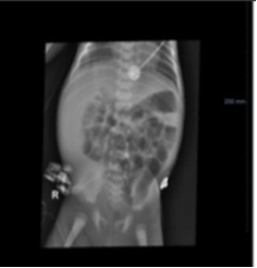
Figure 1. Mild pericholecystic edema and a streak of fluid in Morrison's pouch.
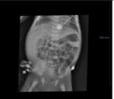
Figure 2. Bilateral mild hydronephrosis and debris in the renal pelvis.
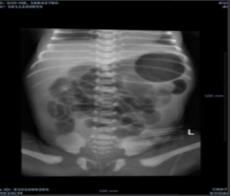
Oblong urinary bladder with irregular walls, multiple sacculations and diverticulae, concerning for neurogenic bladder. No vesicoureteric reflux in filling or micturating phase.
Figure 3. Oblong urinary bladder with irregular walls, multiple sacculations, and diverticulae, concerning for neurogenic bladder; no vesicoureteric reflux was demonstrated during the filling or micturating phase.
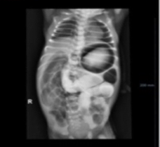
Figure 4. Normal location of the duodenojejunal flexure with no evidence of malrotation; slow transit of contrast with moderately dilated, partially opacified jejunal loops and dilatation of the remaining small-bowel loops suggesting distal obstruction.

3. Result
The two sibling cases demonstrated severe and heterogeneous urinary tract disease. Case 1 was a preterm male neonate with antenatal bilateral hydronephrosis who developed urinary retention, high-grade bilateral vesicoureteral reflux, neurogenic bladder, Burkholderia cepacia sepsis, thrombocytopenia, electrolyte abnormalities, hyperammonemia, and respiratory distress. Case 2 was a female sibling with antenatal megacystis and bilateral hydronephroureter who developed recurrent infections, feeding intolerance, meningitis, and progressive renal failure, ultimately resulting in death at 3.9 years of age.
4. Discussion
Prune Belly Syndrome (PBS), also known as Eagle–Barrett syndrome, is a rare congenital disorder characterized by the classic triad of deficient abdominal wall musculature, urinary tract anomalies, and bilateral cryptorchidism in males. Its incidence is estimated at 1 in 30,000–40,000 live births, with a male predominance. The severity of clinical manifestations can vary, ranging from mild urinary tract dilation to life-threatening renal failure in infancy.
| [11] | Pomajzl AJ, Sankararaman S. Prune Belly Syndrome. In: StatPearls. Treasure Island (FL): StatPearls Publishing; August 8, 2023. |
[11]
In our report, we describe two siblings affected by PBS (or pseudo-PBS), both presenting with severe urinary tract involvement. The first case, a preterm male neonate, had antenatally detected bilateral hydronephrosis and postnatally developed recurrent vomiting, urinary retention, gross vesicoureteral reflux (VUR), and a neurogenic bladder confirmed on MCUG. His course was complicated by hyperammonemia, sepsis due to Burkholderia cepacia, thrombocytopenia, and electrolyte disturbances, requiring prolonged NICU stay. The second case, a female sibling, was diagnosed earlier in life with pseudo-PBS and similarly developed severe urinary tract dilation, sepsis, feeding intolerance, meningitis, and ultimately renal failure, leading to death at 3.9 years of age.
4.1. Comparison with Literature
Our cases highlight several noteworthy aspects. First, the familial clustering of PBS is uncommon but has been reported, suggesting a possible genetic predisposition. Autosomal recessive inheritance has been proposed in some studies, with mutations involving genes such as CHRM3 and HPSE2. The recurrence in siblings, as seen here, underscores the importance of genetic counseling and consideration of molecular testing. Second, while PBS is classically described in males, a pseudo-PBS phenotype may occur in females, often with less severe abdominal wall involvement but equally significant urinary tract disease.
| [3] | Somasundaran, A., Sandra K. Unni, & Naseema Beevi Ahmed Khan. (2024). An interesting case report on obstructed labour with prune belly syndrome. International Journal of Reproduction, Contraception, Obstetrics and Gynecology, 13(9), 2520–2522. https://doi.org/10.18203/2320-1770.ijrcog20242513 |
| [4] | Rupshikha, D., Phukon, M., Kusre, G., Satyanarayana, N., & Sunitha, P. (2012). Prune belly syndrome – A case report. Journal of College of Medical Sciences-Nepal, 7(3), 48–52.
https://doi.org/10.3126/jcmsn.v7i3.6709 |
| [5] | Demisse, A. G., Berhanu, A. & Tadesse, T. Unusual presentation of prune belly syndrome: a case report. J Med Case Reports 11, 337 (2017). https://doi.org/10.1186/s13256-017-1487-9 |
| [8] | Kamoun D, Ben Hamed A, Abdenadher A, Kolsi N, Bouraoui A, Henteti N. The challenging diagnosis and management of the prune belly syndrome: A case report. SAGE Open Medical Case Reports. 2024; 12.
http://doi.org/10.1177/2050313X241258850 |
| [9] | Clince, D.., M. G, D., Ramesh, D., &. S, D. (2022). A Case Report On Prune Belly Syndrome: Pharmaceutical Science-Pharmacy Practice. International Journal of Life Science and Pharma Research, 12(5), L178-P181.
https://doi.org/10.22376/ijpbs/lpr.2022.12.5.L178-181 |
| [14] | De Bernardo G, Giordano M, De Brasi D, Esposito F, De Santis R, Sordino D. Pseudo Prune Belly syndrome: a case report with unilateral abdominal defect. Radiol Case Rep. 2019; 14(8): 941-945. Published 2019 May 29.
https://doi.org/10.1016/j.radcr.2019.05.019 |
| [15] | Singh, Aditya Pratap; Mathur, Vinay; Tanger, Ramesh; Gupta, Arun Kumar. Partial Prune Belly Syndrome: A Rare Case Report. Medical Journal of Dr. D. Y. Patil University 10(5): p 470-472, Sep–Oct 2017. |
https://doi.org/10.4103/MJDRDYPU.MJDRDYPU_280_16 |
[3-5, 8, 9, 14, 15]
4.2. Renal Involvement and Prognosis
Renal dysplasia and progressive renal impairment are the most critical determinants of prognosis in PBS. In case 1, bilateral high-grade VUR with neurogenic bladder placed the child at high risk of chronic kidney disease. Similarly, in case 2, recurrent urinary tract infections (UTIs) and obstructive uropathy led to progressive renal failure. Literature indicates that up to 30–40% of PBS patients progress to end-stage renal disease (ESRD), and early recognition with aggressive urological management is essential to delay this outcome.
| [12] | Cembraneli PN, Cavalcante JBDF, Cavalcante RBDF, Ambrogi G, Cavalcante JEDS. Renal complication due to prune belly syndrome: A case report. Int J Case Rep Images 2020; 11: 101097Z01PC2020. |
[12]
4.3. Management Considerations
Standard management of PBS involves a multidisciplinary approach including neonatology, nephrology, urology, and genetics. Supportive care with electrolyte correction, infection control, and bladder decompression is critical in the acute phase. Surgical interventions such as vesicostomy, ureteral reimplantation, or abdominoplasty are often required, tailored to severity. In case 1, vesicostomy was recommended but deferred due to family issues, highlighting the role of parental decision-making in management outcomes. Case 2 underwent vesicostomy but continued to deteriorate, which reflects that surgical interventions may not always halt disease progression, especially when underlying renal dysplasia is severe.
4.4. Lessons Learned
These cases emphasize several key points:
Genetic predisposition – recurrent PBS/pseudo-PBS in siblings suggests a heritable basis, warranting genetic evaluation and counseling for families.
Early antenatal detection – prenatal ultrasound showing megacystis and hydronephrosis should prompt consideration of PBS.
High morbidity and mortality – both cases demonstrated severe complications (sepsis, electrolyte imbalance, hyperammonemia, renal failure), consistent with poor outcomes in severe PBS phenotypes.
Need for long-term follow-up – multidisciplinary follow-up is essential to manage urinary tract function, prevent infections, and monitor renal function.
5. Conclusion
Our experience with these two sibling cases of Prune Belly Syndrome illustrates the heterogeneity of clinical presentation, the challenges of management in resource-limited settings, and the critical role of early detection and genetic counseling. Awareness of familial recurrence may aid in anticipatory guidance, timely interventions, and better prognostication.
6. Recommendations
Antenatal detection of megacystis and hydronephrosis should prompt early evaluation for PBS and related urinary tract anomalies. Infants with suspected PBS require coordinated multidisciplinary care involving neonatology, pediatric urology, nephrology, and other relevant specialties. Families with recurrent or related congenital anomalies should be offered genetic counseling and consideration of genetic testing. Long-term follow-up should include monitoring of urinary tract function, recurrent infection, and renal function.
Abbreviations
PBS | Prune Belly Syndrome |
VUR | Vesicoureteral Reflux |
NICU | Neonatal Intensive Care Unit |
MCUG | Micturating Cystourethrogram |
HFNC | High-Flow Nasal Cannula |
CRP | C-Reactive Protein |
PRBC | Packed Red Blood Cells |
NPO | Nil Per Os |
OG | Orogastric |
CSF | Cerebrospinal Fluid |
ASD | Atrial Septal Defect |
UTI | Urinary Tract Infection |
ESRD | End-Stage Renal Disease |
Author Contributions
Kashif Shahzad: Conceptualization, Data curation, Investigation, Project administration, Writing – original draft
Saima Ali: Data curation, Investigation
Naveera Waseem: Resources, Visualization, Writing – review & editing
Affan Mudassar: Data curation, Writing – original draft
Raghad Mohammad Ahmed Alshantti: Investigation, Writing – original draft
Karmel AlKhaldy: Writing – original draft, Writing – review & editing
Shehla Chaudhary: Writing – review & editing
Masooma: Writing – review & editing
Conflicts of Interest
The authors declare that they have no competing interests.
References
| [1] |
Giorgi, A. (2024, April 22). Prune belly syndrome: What it is and what to expect. Verywell Health.
https://www.verywellhealth.com/prune-belly-syndrome-8622644
|
| [2] |
Alkhamis, W. H., Abdulghani, S. H. & Altaki, A. Challenging diagnosis of prune belly syndrome antenatally: a case report. J Med Case Reports 13, 198 (2019).
https://doi.org/10.1186/s13256-019-2120-x
|
| [3] |
Somasundaran, A., Sandra K. Unni, & Naseema Beevi Ahmed Khan. (2024). An interesting case report on obstructed labour with prune belly syndrome. International Journal of Reproduction, Contraception, Obstetrics and Gynecology, 13(9), 2520–2522.
https://doi.org/10.18203/2320-1770.ijrcog20242513
|
| [4] |
Rupshikha, D., Phukon, M., Kusre, G., Satyanarayana, N., & Sunitha, P. (2012). Prune belly syndrome – A case report. Journal of College of Medical Sciences-Nepal, 7(3), 48–52.
https://doi.org/10.3126/jcmsn.v7i3.6709
|
| [5] |
Demisse, A. G., Berhanu, A. & Tadesse, T. Unusual presentation of prune belly syndrome: a case report. J Med Case Reports 11, 337 (2017).
https://doi.org/10.1186/s13256-017-1487-9
|
| [6] |
Metwalley, K. A., Farghalley, H. S. & Abd-Elsayed, A. A. Prune belly syndrome in an Egyptian infant with Down syndrome: A case report. J Med Case Reports 2, 322 (2008).
https://doi.org/10.1186/1752-1947-2-322
|
| [7] |
Sood, N. Et al. Trans. 2024. Case report of prune belly syndrome: An airway challenge too?. Indian Journal of Case Reports. 8, 9 (Jan. 2024), 291–292.
https://doi.org/10.32677/ijcr.v8i9.3550
|
| [8] |
Kamoun D, Ben Hamed A, Abdenadher A, Kolsi N, Bouraoui A, Henteti N. The challenging diagnosis and management of the prune belly syndrome: A case report. SAGE Open Medical Case Reports. 2024; 12.
http://doi.org/10.1177/2050313X241258850
|
| [9] |
Clince, D.., M. G, D., Ramesh, D., &. S, D. (2022). A Case Report On Prune Belly Syndrome: Pharmaceutical Science-Pharmacy Practice. International Journal of Life Science and Pharma Research, 12(5), L178-P181.
https://doi.org/10.22376/ijpbs/lpr.2022.12.5.L178-181
|
| [10] |
Kheir, A. E. M., Ali, E. M. A., Medani, S. A., & Maaty, H. S. (2017). Prune belly syndrome: A report of 15 cases from Sudan. Sudanese journal of paediatrics, 17(1), 42–48.
|
| [11] |
Pomajzl AJ, Sankararaman S. Prune Belly Syndrome. In: StatPearls. Treasure Island (FL): StatPearls Publishing; August 8, 2023.
|
| [12] |
Cembraneli PN, Cavalcante JBDF, Cavalcante RBDF, Ambrogi G, Cavalcante JEDS. Renal complication due to prune belly syndrome: A case report. Int J Case Rep Images 2020; 11: 101097Z01PC2020.
|
| [13] |
Snider Novitski K, Radulescu A, Jelin EB, Ladd MR, Meier K, Novotny NM. Prune Belly Syndrome. In: Hirschl RR, Powell DD, Waldhausen JJ, eds. Pediatric Surgery NaT. American Pediatric Surgical Association; 2023.
https://www.pedsurglibrary.com/apsa/view/Pediatric-Surgery-NaT/829694/all/Prune_Belly_Syndrome
Accessed August 25, 2025
|
| [14] |
De Bernardo G, Giordano M, De Brasi D, Esposito F, De Santis R, Sordino D. Pseudo Prune Belly syndrome: a case report with unilateral abdominal defect. Radiol Case Rep. 2019; 14(8): 941-945. Published 2019 May 29.
https://doi.org/10.1016/j.radcr.2019.05.019
|
| [15] |
Singh, Aditya Pratap; Mathur, Vinay; Tanger, Ramesh; Gupta, Arun Kumar. Partial Prune Belly Syndrome: A Rare Case Report. Medical Journal of Dr. D. Y. Patil University 10(5): p 470-472, Sep–Oct 2017. |
https://doi.org/10.4103/MJDRDYPU.MJDRDYPU_280_16
|
Cite This Article
-
APA Style
Shahzad, K., Ali, S., Waseem, N., Mudassar, A., Alshantti, R. M. A., et al. (2026). Prune Belly Syndrome: A Case Series and Literature Review. Science Discovery Pediatrics, 1(1), 13-18. https://doi.org/10.11648/j.sdpediatr.20260101.12
Copy
|
Download
ACS Style
Shahzad, K.; Ali, S.; Waseem, N.; Mudassar, A.; Alshantti, R. M. A., et al. Prune Belly Syndrome: A Case Series and Literature Review. Sci. Discov. Pediatr. 2026, 1(1), 13-18. doi: 10.11648/j.sdpediatr.20260101.12
Copy
|
Download
AMA Style
Shahzad K, Ali S, Waseem N, Mudassar A, Alshantti RMA, et al. Prune Belly Syndrome: A Case Series and Literature Review. Sci Discov Pediatr. 2026;1(1):13-18. doi: 10.11648/j.sdpediatr.20260101.12
Copy
|
Download
-
@article{10.11648/j.sdpediatr.20260101.12,
author = {Kashif Shahzad and Saima Ali and Naveera Waseem and Affan Mudassar and Raghad Mohammad Ahmed Alshantti and Karmel AlKhaldy and Shehla Chaudhary and Masooma},
title = {Prune Belly Syndrome: A Case Series and Literature Review},
journal = {Science Discovery Pediatrics},
volume = {1},
number = {1},
pages = {13-18},
doi = {10.11648/j.sdpediatr.20260101.12},
url = {https://doi.org/10.11648/j.sdpediatr.20260101.12},
eprint = {https://article.sciencepublishinggroup.com/pdf/10.11648.j.sdpediatr.20260101.12},
abstract = {Prune Belly Syndrome (PBS) is a rare congenital disorder characterized by the classical triad of deficient abdominal wall musculature, urinary tract anomalies, and bilateral cryptorchidism in males. The condition is associated with significant morbidity due to urinary tract dysfunction, recurrent infections, and potential renal impairment. We report two siblings with PBS or a pseudo-PBS phenotype, highlighting severe urinary tract involvement and a possible familial predisposition. The first case was a preterm male neonate delivered at 34 weeks of gestation by cesarean section after antenatal ultrasonography demonstrated bilateral hydronephrosis. He subsequently developed recurrent vomiting, progressive abdominal distension, urinary retention, high-grade bilateral vesicoureteral reflux, and a neurogenic bladder. His clinical course was complicated by Burkholderia cepacia sepsis, thrombocytopenia, electrolyte disturbances, hyperammonemia, and respiratory distress requiring intensive care. Pediatric urology recommended vesicostomy and genetic evaluation. The second case was a female sibling with a prenatal diagnosis of a distended bladder and bilateral hydronephroureter who had a pseudo-PBS phenotype and developed severe urinary tract disease, recurrent infections, feeding intolerance, meningitis, and progressive renal failure, ultimately resulting in death at 3.9 years of age. These cases emphasize the importance of early recognition of PBS, particularly when antenatal urinary tract abnormalities and a positive family history are present, and highlight the need for multidisciplinary management, genetic counseling, and long-term renal and urological follow-up. A preterm male neonate was delivered at 34 weeks of gestation via cesarean section. Antenatal ultrasonography revealed bilateral hydronephrosis. The patient had a significant family history of a sibling who had died from pseudo-prune belly syndrome. Shortly after birth, he developed recurrent vomiting, progressive abdominal distension, and urinary retention. Radiological investigations demonstrated severe bilateral vesicoureteral reflux and a neurogenic bladder, confirming the diagnosis of PBS. During hospitalization, the infant developed sepsis caused by Burkholderia cepacia, accompanied by electrolyte disturbances and hyperammonemia, necessitating intensive care management. Treatment included broad-spectrum antibiotics, blood transfusions, ventilatory support, and close monitoring. Pediatric urology consultation recommended vesicostomy to relieve urinary obstruction and genetic testing to evaluate the possible familial basis of the disorder. This case highlights the importance of early recognition of PBS, particularly in neonates with antenatal urinary tract abnormalities and a positive family history. The occurrence of severe complications, including sepsis and metabolic derangements, underscores the need for prompt multidisciplinary intervention. Genetic counseling and evaluation should be considered in families with recurrent or related congenital anomalies to facilitate early diagnosis, guide management, and improve long-term outcomes.},
year = {2026}
}
Copy
|
Download
-
TY - JOUR
T1 - Prune Belly Syndrome: A Case Series and Literature Review
AU - Kashif Shahzad
AU - Saima Ali
AU - Naveera Waseem
AU - Affan Mudassar
AU - Raghad Mohammad Ahmed Alshantti
AU - Karmel AlKhaldy
AU - Shehla Chaudhary
AU - Masooma
Y1 - 2026/10/21
PY - 2026
N1 - https://doi.org/10.11648/j.sdpediatr.20260101.12
DO - 10.11648/j.sdpediatr.20260101.12
T2 - Science Discovery Pediatrics
JF - Science Discovery Pediatrics
JO - Science Discovery Pediatrics
SP - 13
EP - 18
PB - Science Publishing Group
UR - https://doi.org/10.11648/j.sdpediatr.20260101.12
AB - Prune Belly Syndrome (PBS) is a rare congenital disorder characterized by the classical triad of deficient abdominal wall musculature, urinary tract anomalies, and bilateral cryptorchidism in males. The condition is associated with significant morbidity due to urinary tract dysfunction, recurrent infections, and potential renal impairment. We report two siblings with PBS or a pseudo-PBS phenotype, highlighting severe urinary tract involvement and a possible familial predisposition. The first case was a preterm male neonate delivered at 34 weeks of gestation by cesarean section after antenatal ultrasonography demonstrated bilateral hydronephrosis. He subsequently developed recurrent vomiting, progressive abdominal distension, urinary retention, high-grade bilateral vesicoureteral reflux, and a neurogenic bladder. His clinical course was complicated by Burkholderia cepacia sepsis, thrombocytopenia, electrolyte disturbances, hyperammonemia, and respiratory distress requiring intensive care. Pediatric urology recommended vesicostomy and genetic evaluation. The second case was a female sibling with a prenatal diagnosis of a distended bladder and bilateral hydronephroureter who had a pseudo-PBS phenotype and developed severe urinary tract disease, recurrent infections, feeding intolerance, meningitis, and progressive renal failure, ultimately resulting in death at 3.9 years of age. These cases emphasize the importance of early recognition of PBS, particularly when antenatal urinary tract abnormalities and a positive family history are present, and highlight the need for multidisciplinary management, genetic counseling, and long-term renal and urological follow-up. A preterm male neonate was delivered at 34 weeks of gestation via cesarean section. Antenatal ultrasonography revealed bilateral hydronephrosis. The patient had a significant family history of a sibling who had died from pseudo-prune belly syndrome. Shortly after birth, he developed recurrent vomiting, progressive abdominal distension, and urinary retention. Radiological investigations demonstrated severe bilateral vesicoureteral reflux and a neurogenic bladder, confirming the diagnosis of PBS. During hospitalization, the infant developed sepsis caused by Burkholderia cepacia, accompanied by electrolyte disturbances and hyperammonemia, necessitating intensive care management. Treatment included broad-spectrum antibiotics, blood transfusions, ventilatory support, and close monitoring. Pediatric urology consultation recommended vesicostomy to relieve urinary obstruction and genetic testing to evaluate the possible familial basis of the disorder. This case highlights the importance of early recognition of PBS, particularly in neonates with antenatal urinary tract abnormalities and a positive family history. The occurrence of severe complications, including sepsis and metabolic derangements, underscores the need for prompt multidisciplinary intervention. Genetic counseling and evaluation should be considered in families with recurrent or related congenital anomalies to facilitate early diagnosis, guide management, and improve long-term outcomes.
VL - 1
IS - 1
ER -
Copy
|
Download