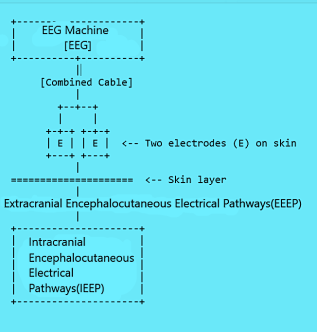
The traditional understanding of electroencephalogram (EEG) genesis and propagation center on the summation of neuronal field potentials transmitted to the scalp via passive volume conduction. While this neurocentric view has dominated EEG interpretation for decades, certain bioelectrical observations—such as the abrupt disappearance of EEG activity following cerebral circulatory arrest—suggest that a more nuanced mechanism may underlie the transmission of brain-derived electrical signals to the body surface. This paper introduces a novel vascular-electrical hypothesis termed Encephalocutaneous Electrical Pathways (EEP), which proposes that specialized, non-neuronal electrical conduits—distinct from classical axonal pathways—exist within both intracranial and extracranial compartments and serve to transmit bioelectrical activity from the brain to the skin surface. EEP comprises two structurally integrated components: an intracranial network of microscopic electrical pathways, hypothesized to propagate bioelectricity independently of synaptic transmission and aligned with perivascular microenvironments, and an extracranial extension of these pathways that follows vascular channels through the meninges and connective tissues to terminate in the skin. These encephalocutaneous terminations constitute the interface through which EEG signals emerge on the scalp. Although closely associated with vascular architecture, EEP structures are not vascular themselves, but anatomically distinct entities that utilize the vascular corridors for spatial orientation. This hypothesis accounts for EEG disappearance following loss of cerebral perfusion and posits a direct, vascular-aligned but non-vascular electrical continuum from brain to skin. If validated, the EEP model would challenge conventional views of EEG signal origin and propagation and establish a foundation for exploring extracranial bioelectric communication. It opens new directions for anatomical mapping, electrophysiological validation, and the development of novel EEG technologies that account for encephalocutaneous bioelectricity.
| Published in | Science Innovation (Volume 13, Issue 4) |
| DOI | 10.11648/j.si.20251304.11 |
| Page(s) | 57-62 |
| Creative Commons |
This is an Open Access article, distributed under the terms of the Creative Commons Attribution 4.0 International License (http://creativecommons.org/licenses/by/4.0/), which permits unrestricted use, distribution and reproduction in any medium or format, provided the original work is properly cited. |
| Copyright |
Copyright © The Author(s), 2025. Published by Science Publishing Group |
Encephalocutaneous Electrical Pathways (EEP), Electroencephalogram (EEG), Bioelectricity, Vascular Channels, Non-Neuronal Electrophysiology, Brain-Skin Bioelectrical Interface
BCI | Brain-Computer Interface |
EEG | Electroencephalogram |
EEP | Encephalocutaneous Electrical Pathways |
EEEP | Extracranial Encephalocutaneous Electrical Pathways |
IEEP | Intracranial Encephalocutaneous Electrical Pathways |
| [1] | Nunez PL, Srinivasan R. Electric fields of the brain: The neurophysics of EEG. 2nd ed. Oxford University Press; 2006. |
| [2] | Iliff JJ, Wang M, Liao Y, et al. A paravascular pathway facilitates CSF flow through the brain parenchyma and the clearance of interstitial solutes. Sci Transl Med. 2012; 4(147): 147ra111. |
| [3] | Nedergaard M, Goldman SA. Glymphatic failure as a final common pathway to dementia. Science. 2020; 370(6512): 50-56. |
| [4] | Mughal A, Hennig GW, Heppner T, Tsoukias NM, Hill-Eubanks D, Nelson MT. Electrocalcium coupling in brain capillaries: Rapidly traveling electrical signals ignite local calcium signals. Proceedings of the National Academy of Sciences. 2024 Dec 17; 121(51): e2415047121. |
| [5] | Hakim MA, Behringer EJ, Nelson MT. K_IR channel regulation of electrical conduction along cerebrovascular endothelium: enhanced modulation during Alzheimer's disease. Microcirculation. 2023; 30(1): e12797. |
| [6] | Buzsáki G, Anastassiou CA, Koch C. The origin of extracellular fields and currents—EEG, ECoG, LFP and spikes. Nat Rev Neurosci. 2012; 13(6): 407-420. |
| [7] | Chiang CC, Shivacharan RS, Durand DM. Slow periodic activity in hippocampal slice propagates via ephaptic coupling. J Physiol. 2018; 596(22): 5513-5529. |
| [8] | Ihne J, Lindström M, Halén J. Astrocytic endfoot calcium waves regulate cerebral blood flow independent of neuronal activation. eLife. 2021; 10: e64311. |
| [9] | Zhang Y, Shao Y, Wang Y, et al. From mechanisms to medicine: neurovascular coupling in cerebrovascular disorders. Cells. 2023; 14(1): 16. |
| [10] | Wardlaw JM, Smith C, Dichgans M. Small vessel disease: mechanisms and clinical implications. Lancet Neurol. 2019; 18(7): 684-696. |
| [11] | Finger S. Origins of Neuroscience: A History of Explorations into Brain Function. Oxford University Press; 1994. |
| [12] | Gross CG. Brain, vision, memory: tales in the history of neuroscience. Trends Neurosci. 1999; 22(1): 27-30. |
| [13] | Clarke E, O'Malley CD. The Human Brain and Spinal Cord: A Historical Study Illustrated by Writings from Antiquity to the Twentieth Century. University of California Press; 1996. |
| [14] | Caton R. The electric currents of the brain. BrMedJ. 1875; 2(765): 278. |
| [15] | Berger H. Über das Elektrenkephalogramm des Menschen. Arch Psychiatr Nervenkr. 1929; 87: 527-570. |
| [16] | Gloor P. Hans Berger on the electroencephalogram of man. Electroencephalogr Clin Neurophysiol Suppl. 1969; 28: 1-350. |
| [17] | Cobb W, Sears TA. A study of the transmission of potentials after hemispherectomy. Electroencephalogr Clin Neurophysiol. 1960; 12(2): 371-83. |
| [18] | Lopes da Silva FH. EEG and MEG: relevance to neuroscience. Neuron. 2013; 80(5): 1112-1128. |
| [19] | Libenson MH. Clinical EEG monitoring after cardiac arrest. Intensive Care Med. 2022; 48(3): 389-414. |
| [20] | Longden TA, Nelson MT. Regulation of cerebral blood flow by endothelial electrical signaling. Glia. 2021; 69(5): 1048-1063. |
| [21] | Guerra G, Lucariello A, Perna A, Botta L, DeLuca A, Moccia F. The Role of Endothelial Ca²⁺ Signaling in Neurovascular Coupling: A View from the Lumen. Int J Mol Sci. 2018; 19(4): 938. |
| [22] | Bazargani N, Attwell D. Astrocyte calcium signaling: the third wave. Nat Neurosci. 2016; 19(2): 182-189. |
| [23] | Frazier AE, Thompson RM, Halliday GM, Shepherd RK, Dottori M. Non-neuronal cells in the neurovascular unit: key roles in health and neurodegenerative disease. Front Neurosci. 2021; 15: 664313. |
| [24] | BA, Newman EA. Astrocyte regulation of blood flow in the brain. Cold Spring Harb Perspect Biol. 2015; 7(5): a020388. |
| [25] | Greger B, Halgren E, Schalk G. The neurophysiology of brain-computer interfaces: past, present, and future. IEEE Trans Biomed Eng. 2020; 67(11): 3093-3105. |
| [26] | Fukada E, Yasuda I. On the piezoelectric effect of bone. J Phys Soc Jpn. 1957; 12(10): 1158-1162. |
| [27] | Claassen J, Hirsch LJ, Kreiter KT, Du EY, Connolly ES, Mayer SA. Quantitative continuous EEG for detecting delayed cerebral ischemia in subarachnoid hemorrhage. Clin Neurophysiol. 2004; 115(12): 2699-2710. |
| [28] | Shih AY, Driscoll JD, Drew PJ, Nishimura N, Schaffer CB, Kleinfeld D. Two-photon microscopy as a tool to study blood flow and neurovascular coupling in the rodent brain. J Cereb Blood Flow Metab. 2012; 32(7): 1277-309 |
| [29] | Wang XJ. Macroscopic gradients of synaptic excitation and inhibition in the neocortex. Nat Rev Neurosci. 2020; 21(3): 169-178. |
| [30] | Araque A, Carmignoto G, Haydon PG, Oliet SHR, Robitaille R, Volterra A. Gliotransmitters travel in time and space. Neuron. 2014; 81(4): 728-739. |
| [31] | Kisler K, Nelson AR, Montagne A, Zlokovic BV. Cerebral blood flow regulation and neurovascular dysfunction in Alzheimer disease. Nat Rev Neurosci. 2017; 18(7): 419-434. |
| [32] | Thomsen K, Piilgaard H, Gjedde A, Bonvento G, Lauritzen M. Principal cell spiking, postsynaptic excitation, and oxygen consumption in the rat cerebellum. J Neurosci. 2009; 29(7): 2185-2195. |
| [33] | Rash JE. Molecular disruptions of the panglial syncytium block potassium siphoning and axonal saltatory conduction: pertinence to neuromyelitis optica and other demyelinating diseases of the central nervous system. Neuroscience. 2010; 168(4): 982-1008. |
| [34] | Hallez H, Vanrumste B, Van Hese P, D'Asseler Y, Lemahieu I, Van de Walle R. Dipole estimation errors due to differences in modeling anisotropic conductivities in realistic head models for EEG source analysis. Phys Med Biol. 2008; 53(7): 1877-1894. |
| [35] | Johnson EL, Young ML, Moore SK, et al. Long-term electroencephalography monitoring in critically ill patients: practical considerations and clinical relevance. J Clin Neurophysiol. 2021; 38(1): 3-14. |
| [36] | Liao LD, Wang IJ, Chen SF, Chang JY, Lin CT. Design, fabrication and experimental validation of a novel dry-contact sensor for measuring electroencephalography signals without skin preparation. Sensors (Basel). 2011; 11(6): 5819-5834. |
APA Style
Ogunlade, O. (2025). Beyond Volume Conduction: The Encephalocutaneous Electrical Pathways (EEP) Hypothesis for Vascular-Aligned, Non-Neuronal EEG Genesis and Propagation. Science Innovation, 13(4), 57-62. https://doi.org/10.11648/j.si.20251304.11
ACS Style
Ogunlade, O. Beyond Volume Conduction: The Encephalocutaneous Electrical Pathways (EEP) Hypothesis for Vascular-Aligned, Non-Neuronal EEG Genesis and Propagation. Sci. Innov. 2025, 13(4), 57-62. doi: 10.11648/j.si.20251304.11
AMA Style
Ogunlade O. Beyond Volume Conduction: The Encephalocutaneous Electrical Pathways (EEP) Hypothesis for Vascular-Aligned, Non-Neuronal EEG Genesis and Propagation. Sci Innov. 2025;13(4):57-62. doi: 10.11648/j.si.20251304.11
@article{10.11648/j.si.20251304.11,
  author = {Oluwadare Ogunlade},
  title = {Beyond Volume Conduction: The Encephalocutaneous Electrical Pathways (EEP) Hypothesis for Vascular-Aligned, Non-Neuronal EEG Genesis and Propagation
},
  journal = {Science Innovation},
  volume = {13},
  number = {4},
  pages = {57-62},
  doi = {10.11648/j.si.20251304.11},
  url = {https://doi.org/10.11648/j.si.20251304.11},
  eprint = {https://article.sciencepublishinggroup.com/pdf/10.11648.j.si.20251304.11},
  abstract = {The traditional understanding of electroencephalogram (EEG) genesis and propagation center on the summation of neuronal field potentials transmitted to the scalp via passive volume conduction. While this neurocentric view has dominated EEG interpretation for decades, certain bioelectrical observations—such as the abrupt disappearance of EEG activity following cerebral circulatory arrest—suggest that a more nuanced mechanism may underlie the transmission of brain-derived electrical signals to the body surface. This paper introduces a novel vascular-electrical hypothesis termed Encephalocutaneous Electrical Pathways (EEP), which proposes that specialized, non-neuronal electrical conduits—distinct from classical axonal pathways—exist within both intracranial and extracranial compartments and serve to transmit bioelectrical activity from the brain to the skin surface. EEP comprises two structurally integrated components: an intracranial network of microscopic electrical pathways, hypothesized to propagate bioelectricity independently of synaptic transmission and aligned with perivascular microenvironments, and an extracranial extension of these pathways that follows vascular channels through the meninges and connective tissues to terminate in the skin. These encephalocutaneous terminations constitute the interface through which EEG signals emerge on the scalp. Although closely associated with vascular architecture, EEP structures are not vascular themselves, but anatomically distinct entities that utilize the vascular corridors for spatial orientation. This hypothesis accounts for EEG disappearance following loss of cerebral perfusion and posits a direct, vascular-aligned but non-vascular electrical continuum from brain to skin. If validated, the EEP model would challenge conventional views of EEG signal origin and propagation and establish a foundation for exploring extracranial bioelectric communication. It opens new directions for anatomical mapping, electrophysiological validation, and the development of novel EEG technologies that account for encephalocutaneous bioelectricity.},
 year = {2025}
}
TY  - JOUR
T1  - Beyond Volume Conduction: The Encephalocutaneous Electrical Pathways (EEP) Hypothesis for Vascular-Aligned, Non-Neuronal EEG Genesis and Propagation

AU  - Oluwadare Ogunlade
Y1  - 2025/07/23
PY  - 2025
N1  - https://doi.org/10.11648/j.si.20251304.11
DO  - 10.11648/j.si.20251304.11
T2  - Science Innovation
JF  - Science Innovation
JO  - Science Innovation
SP  - 57
EP  - 62
PB  - Science Publishing Group
SN  - 2328-787X
UR  - https://doi.org/10.11648/j.si.20251304.11
AB  - The traditional understanding of electroencephalogram (EEG) genesis and propagation center on the summation of neuronal field potentials transmitted to the scalp via passive volume conduction. While this neurocentric view has dominated EEG interpretation for decades, certain bioelectrical observations—such as the abrupt disappearance of EEG activity following cerebral circulatory arrest—suggest that a more nuanced mechanism may underlie the transmission of brain-derived electrical signals to the body surface. This paper introduces a novel vascular-electrical hypothesis termed Encephalocutaneous Electrical Pathways (EEP), which proposes that specialized, non-neuronal electrical conduits—distinct from classical axonal pathways—exist within both intracranial and extracranial compartments and serve to transmit bioelectrical activity from the brain to the skin surface. EEP comprises two structurally integrated components: an intracranial network of microscopic electrical pathways, hypothesized to propagate bioelectricity independently of synaptic transmission and aligned with perivascular microenvironments, and an extracranial extension of these pathways that follows vascular channels through the meninges and connective tissues to terminate in the skin. These encephalocutaneous terminations constitute the interface through which EEG signals emerge on the scalp. Although closely associated with vascular architecture, EEP structures are not vascular themselves, but anatomically distinct entities that utilize the vascular corridors for spatial orientation. This hypothesis accounts for EEG disappearance following loss of cerebral perfusion and posits a direct, vascular-aligned but non-vascular electrical continuum from brain to skin. If validated, the EEP model would challenge conventional views of EEG signal origin and propagation and establish a foundation for exploring extracranial bioelectric communication. It opens new directions for anatomical mapping, electrophysiological validation, and the development of novel EEG technologies that account for encephalocutaneous bioelectricity.
VL  - 13
IS  - 4
ER  -
Department of Physiological Sciences, Obafemi Awolowo University, Ile-Ife, Nigeria
Research Fields: Physiological Sciences, Medicine (cardiology), Maturology and Lenism
Information